
Functional examination of the knee
Functional examination of the knee
Interesting to know before examining the patient
Pain felt at the knee mostly suggests a local lesion. Exceptionally, the pain is referred : from an L2/L3-structure if it is felt at the front of the knee and from an S1/S2-structure if the pain is felt at the back of the knee or from an L4/L5 structure if the pain is lateral from the knee.
I refer to the specific rules of soft tissue referred pain which are described in another chapter.
There is a particularity at the knee, which is rather uncommon at e.g. the hip- or shoulder joint. Some hours after an articular or ligamentous injury of some severity a post-traumatic capsular reaction develops. The clinical image of a traumatic arthritis will dominate the clinical pattern, as from the next day, resulting in a capsular pattern and swelling.
Sprains of ligaments which form an integral part of the capsule itself (e.g. anterior talofibular ligament or the medial collateral ligament) cause a secondary capsulitis and hence a non-capsular pattern is superimposed on the capsular pattern
As a consequence the initial, non-capsular lesion will be more difficult to find. Therefore, the history at the knee is much more important than at the other peripheral joints.
The capsular pattern of the knee is a limitation of flexion and a smaller limitation of extension.
>>> However, in literature there seems to be some confusion:
We need to make a distinction between a minor arthrosis and a clear arthritis.
In case of a minor arthrosis indeed it is possible that we don’t find the capsular pattern (flexion is limited but extension not yet) and that just one movement is (slightly) limited instead of two.
In case of an acute arthritis, we certainly will find a capsular pattern.
The main cause of a non-capsular pattern is an internal derangement from a loose body or a damaged meniscus.
The specific history tells you more
Age and profession
A similar history can suggest different pathologies, according to the patient’s age.
For example : the patient describes the history of an internal derangement (twinges, locking, giving way). Depending on the age, different pathologies may be present.
- If the patient is 25 years old, a torn meniscus is the first thing we think of
- if he is 65 years old, it sounds more like a loose body in an osteoarthrotic joint
- At the age of 15 , an osteochondritis dissecans (loose body with a bony nucleus) is likely.
Onset of symptoms : spontaneous or traumatic ?
In case of a traumatic event, it is interesting to know which forces were acting upon the knee? In which position was de knee ?
- A flexion-lateral rotation strain --> first think of medial coronary ligament, in a more severe lesion we think of MCL and menisci
- A flexion-medial rotation strain --> first think of lateral coronary ligament
- A valgus strain --> medial collateral ligament ?
- A varus strain --> lateral collateral ligament?
- An extension trauma --> Cruciate ligaments ?
Was there any sudden giving way, or locking ?
A sudden locking in flexion after trauma, without spontaneous unlocking, suggests a severe meniscal lesion (bucket handle tear). Temporary locking in extension, with spontaneous unlocking rather suggests a loose body or a minor meniscal lesion.
Site of the pain : at one side, inside the knee, diffuse, alternating ?
Pain inside the knee is typical for a lesion of a cruciate ligament ; diffuse pain suggests arthritis. An alternating or shifting pain points in the direction of an internal derangement (loose body or menisc).
Did the joint swell, immediately or after some time ?
Immediate swelling after trauma suggests haemarthrosis, swelling after some time indicates synovial fluid related to the post-traumatic capsular reaction. In an overuse tendinitis or tendinosis we don’t expect swelling.
Was there any loss of function, how soon ?
Immediate loss of function after trauma suggests a torn meniscus ; loss of function, appearing after a few hours, is part of the post-traumatic capsular reaction and a ligamentous lesion is then more probable.
What was the evolution of the symptoms ?
- Has the pain spread, where to ?
- Have there been any recurrences ?
- What is the effect of going up and down stairs ? Is the one more troublesome than the other ?
A patient suffering from a loose body in an osteoarthrotic joint has a certain apprehension of going down stairs, feeling insecure. (Therefore, an interesting test after successful manipulation for a loose body is to request the patient to go downstairs : if successful,his insecurity will be clearly gone.)
Are there any twinges ?
Does the knee click or grate ?
Have there been previous treatments, which were the results ?
Have there been other joints affected ?
The combination of twinges, locking and a feeling of giving way always points in the direction of an internal derangement.
How to perfom a basic functional examination
Palpation for local heat
 Palpation for local heath
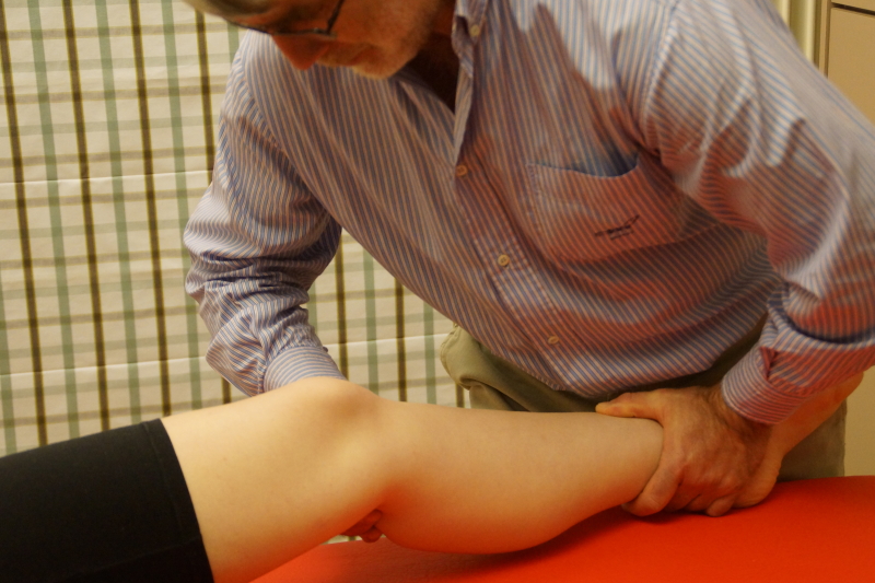
Passive flexion
 passive knee flexion
The end-feel is extra-articular or elastic.
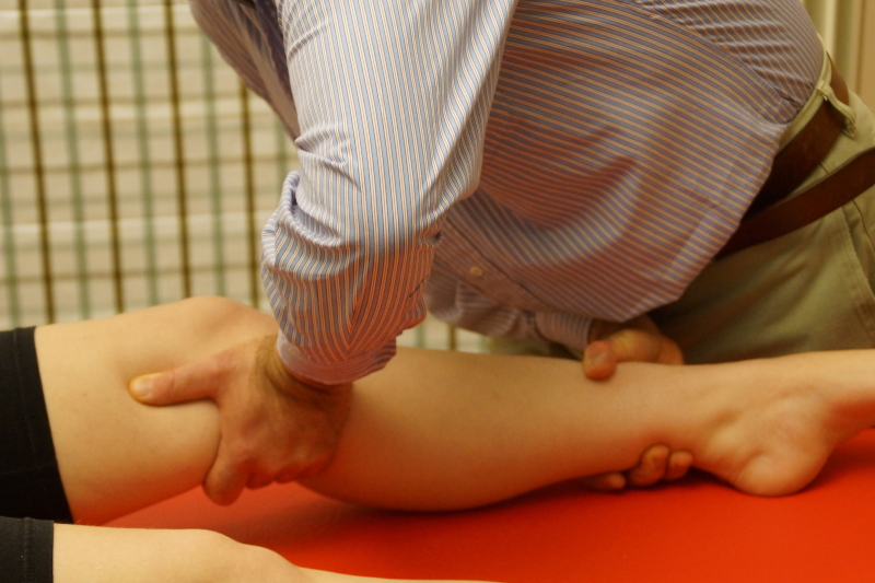
Passive extension
 passive knee extension
End feel is rather hard, because of the tension in the cruciate ligaments.
After these two articular tests a capsular pattern may be found : flexion is more limited than extension.
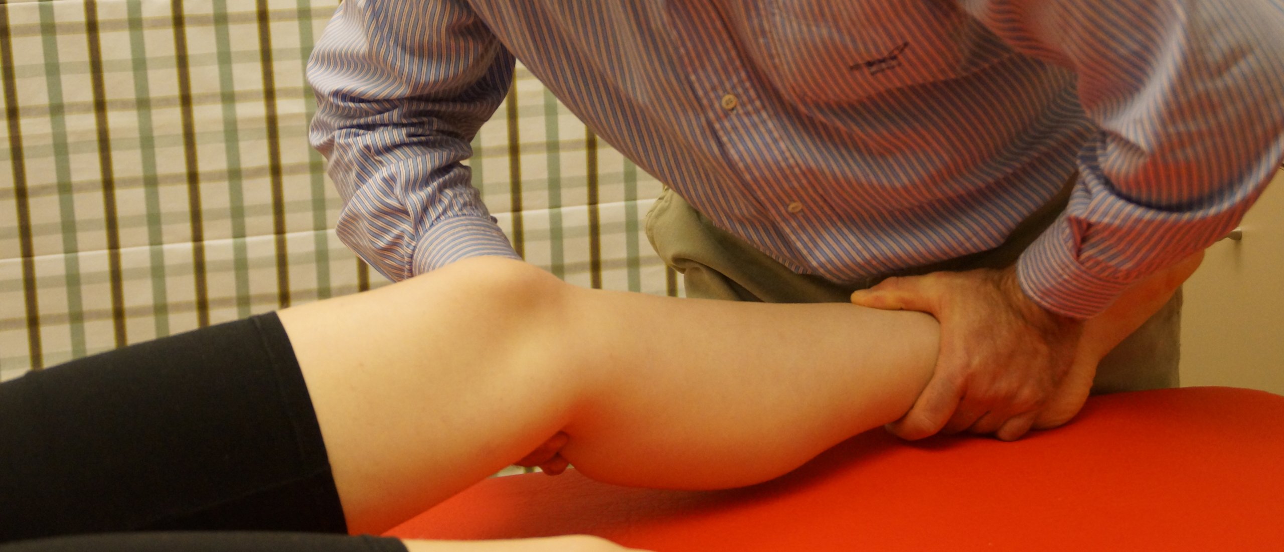
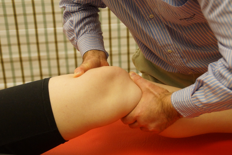
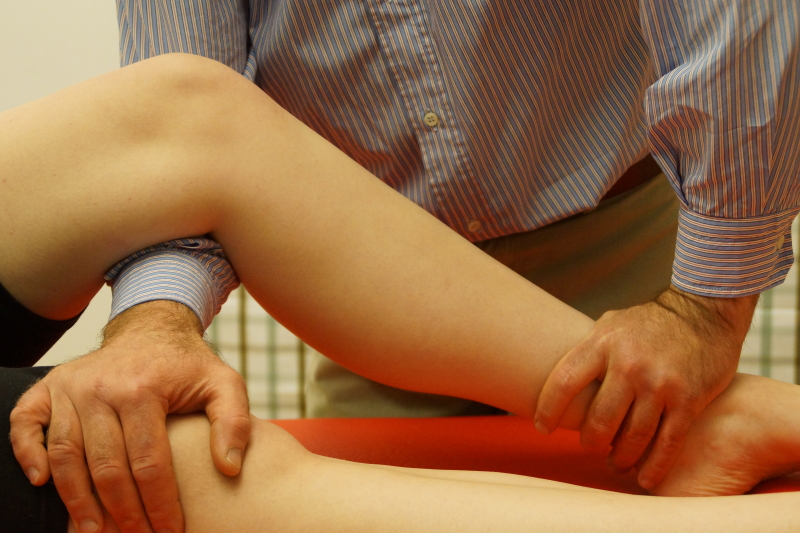
Passive lateral rotation
 passive lateral rotation
The ankle joint is somewhat blocked in dorsiflexion and we use the medial boarder of the foot as a lever ; go to the end of the movement and then slightly provoke. This the main test for the medial coronary ligament.
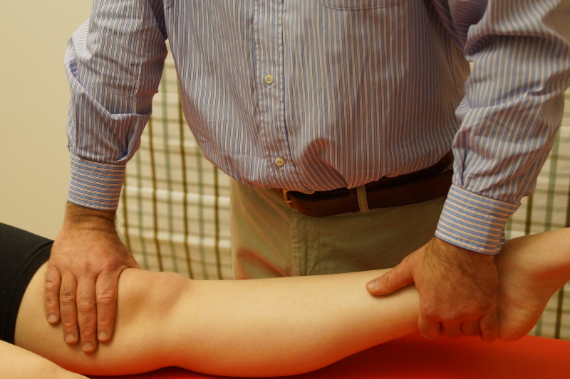
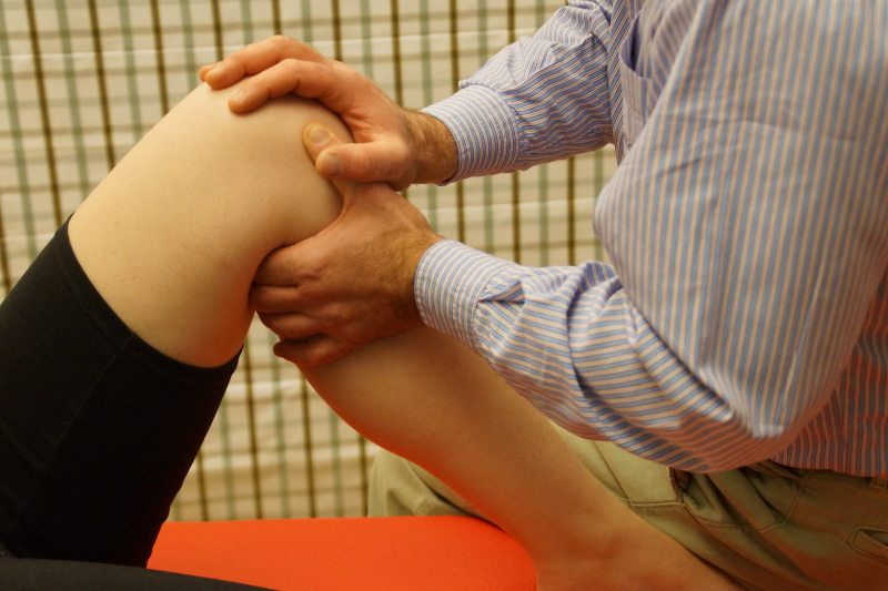
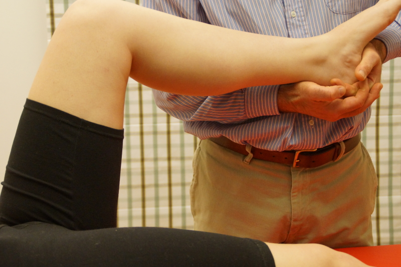
Passive medial rotation
 passive medial rotation
This the main test for the lateral coronary ligament.
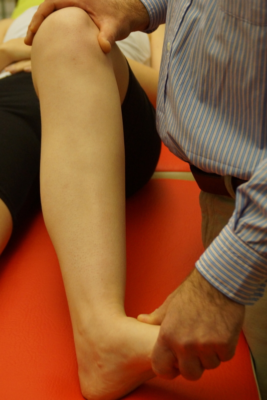
Passive valgus strain
 passive valgus strain
Take up the slack and provoke towards valgus. Keep in mind that the knee is in slight flexion postion, otherwise we can’t perform the test.
This is the main test for the MCL.
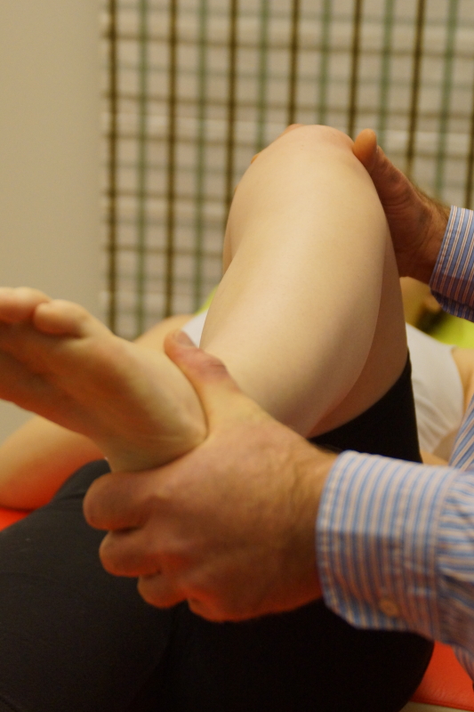
Passive varus strain
 passive varus strain
Main test for the LCL.
Lachman test
 Lachman test
We need to compare both sides and welook for hypermobility, pointing on the direction of an anterior cruciate ligament lesion.
As an alternative we can also use Lelli’s test.
Posterior drawer test
 Posterior drawer test
While pushing on the tibia in a posterior direction, we palpate with the thumb in the joint line. In normal circumstances no movement is felt.
If mobility is felt, most likely there is a posterior cruciate ligament lesion.
Resisted extension
 Resisted extension
Main test for the quadriceps.
Resisted flexion
 resisted flexion
Test for the hamstrings, biceps femoris, pes anserinus and popliteus.
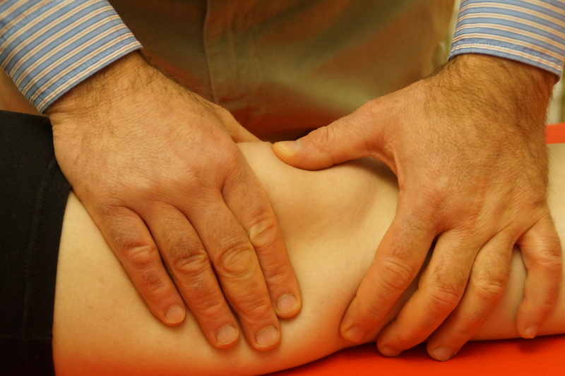
Palpation
- Warmth, fluid, synovial thickening
Warmth and fluid suggest an articular or ligamentous lesion in an active stage.
The presence of fluid can be elicited by two tests : one for a large quantity of fluid in the joint (the patellar tap) and one for a minor effusion.
Synovial thickening suggests a rheumatoid type of arthritis. It is palpated at the femoral condyles.
 Palpation for swelling (1)
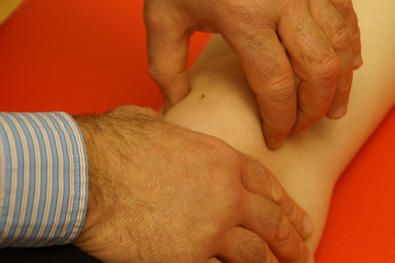 Palpation for swelling (2)
2. Tenderness in the affected structure
3. Other findings : cyst, osteophyte, crepitus
A series of complementary mensical tests are described in literature, but all of them seem to have a rather limited validity.
The most useful meniscal test seems to be following:
Ask the patient to stand on one leg, slightly flex the knee, and then perform a pivoting movement. We try to provoke the specific symptoms the patient described in the history.
Place comment