Shoulder functional examination
Shoulder non-capsular pattern, the diagnostic options

When the patient suffers from a tendinitis/tendinosis, then the history will provide us some basic information. In case of an arthritis however, then the history becomes much more important.
It will help us to determine which kind of arthritis it is (traumatic ?,…) and will also reveal the stage of the arthritis. How acute or chronic is it?
This is very important information since the treatment strategy in each stage is quite different.
We ask the standard questions :
Focus more specifically on following questions:
Are the patient’s symptoms the result of a local lesion or is the pain referred from e.g. the cervical spine ?
Remember that shoulder structures have a C5-origin and thus will refer pain somewhere in the C5-dermatome. The AC-joint will refer pain in the C4-dermatome.
Of course the specific history will mostly provide you enough information to decide if it is a shoulder or a cervical problem.
(in case there is any doubt: also check some cervical functional tests and see whether or not you can provoke the patient’s symptoms by doing those tests).
A capsular pattern found after injury or overuse most likely suggests a traumatic arthritis. Keep in mind other types of arthritis do exist.
A traumatic arthritis apparently does not occur under the age of 40-45 y. This is a purely empirical finding for which we don’t have solid scientific proof yet.
It’s not only interesting to know whether the lesion is acute or chronic.
Some lesions also have a spontaneous evolution, and that might have a consequence on the therapy level:
Example :
Depeding on in which phase of the spontaneous evolution the lesion is, the treatment will be different. (I refer to other chapters for more details).
If so, then we have to consider disorders such as rheumatoid arthritis., psoriatic, lupus erythematosus,…
If the patient had a breast surgery e.g. 3 years ago and now she/he presents with a shoulder problem, then be aware of inherent unlikelyhoods.
Stage I arthritis :
This is an arthritis “light”.
There is a favourable answer on the three questions :
Stage III arthritis :
This is an acute arthritis.
There is a non-favourable answer on the three questions :
Stage II arthritis :
Depending on the stage, the treatment procedure will be different.
Sometimes symptoms in the shoulder area are referred from the cervical spine.
If necessary, we perform a preliminary cervical examination for differential diagnostic purposes.
If cervical tests cleary influence the patient’s symptoms then we have a positive link and we perform the complete cervical examination. If cervical tests are negative, we proceed with the shoulder examination.
The challenge is to collect as much information as possible by using as less tests as possible. There is no need for excessive testing. If necessary, some complementary tests (see film below) may be useful.
All tests need to be performed bilaterally.
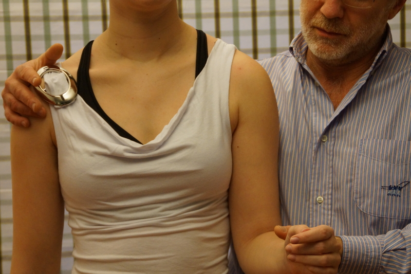
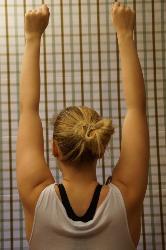 Active elevation
We assess pain and range of motion (ROM). Although this test does not enable us to differentiate between a lesion of an inert or a contractile structure, it gives us a good idea of the patient’s willingness to move.
This is not a glenohumeral test but rather an aspecific shoulder girdle test.
We distinguish three components :
Beware of some inherent unlikelihoods :
even with a completely ankylosed shoulder joint, when muscles and nerves function normally, there still has to be some 60° of arm elevation, due to the intact scapular rotation.
Also keep in mind that the range on passive elevation is always bigger than that on passive glenohumeral abduction.
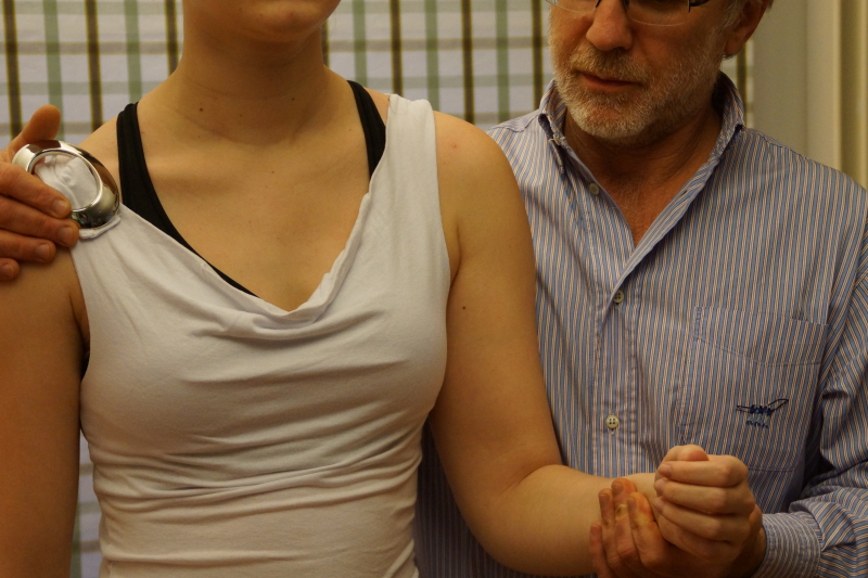
 Passive elevation
We assess pain, ROM and end-feel (elastic).
Grasp the elbow, not the forearm, for a more precise execution of the test.
If the shoulder is quite mobile, ask the patient to perform an active neck flexion to create some space for the movement.
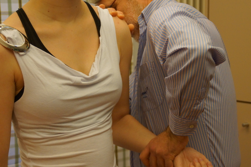
 Looking for a painful arc
Ask the patient to do an elevation movement, slowly, and he has to tell us what he feels, where and when.
We may not miss a painful arc in the shoulder, since mostly this is an important localizing sign.
So, we are looking for a painful movement section, inbetween two painfree sections.
Indicating something is squeezed in between humerus and acromion. (more details will be discussed in another chapter).
If the arc is so severe that the patient is unable to lift his arm beyond the horizontal without help of the other arm (i.e. a passive painful arc), severe calcification in a tendon or a bursa is suspected. This is an empirical finding.
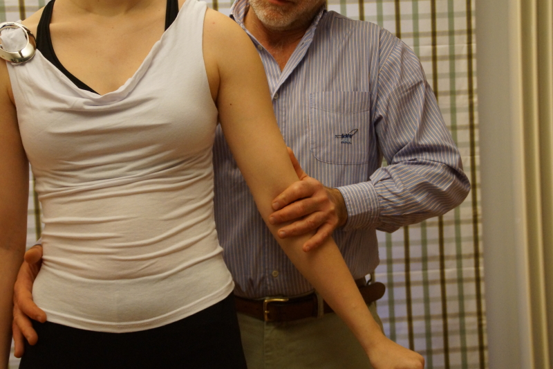
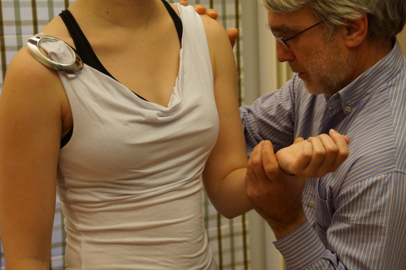
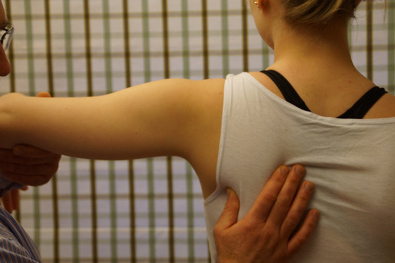 Passive glenohumeral abduction
The thumb fixes the inferior angle of the scapula, the arm is raised passively untill we feel the end of movement. The scapula remains fixed. We assess the ROM (normally 90°).
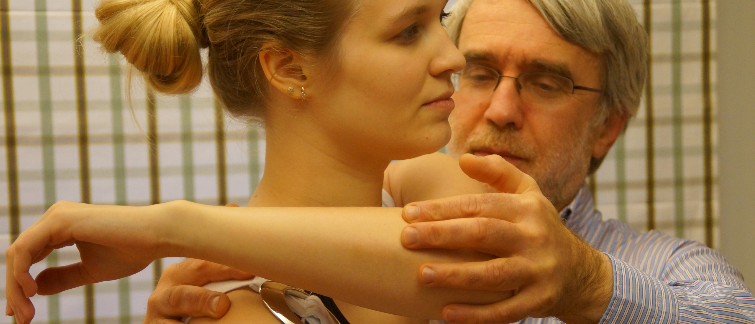
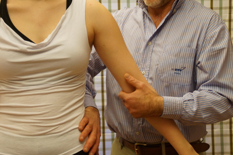
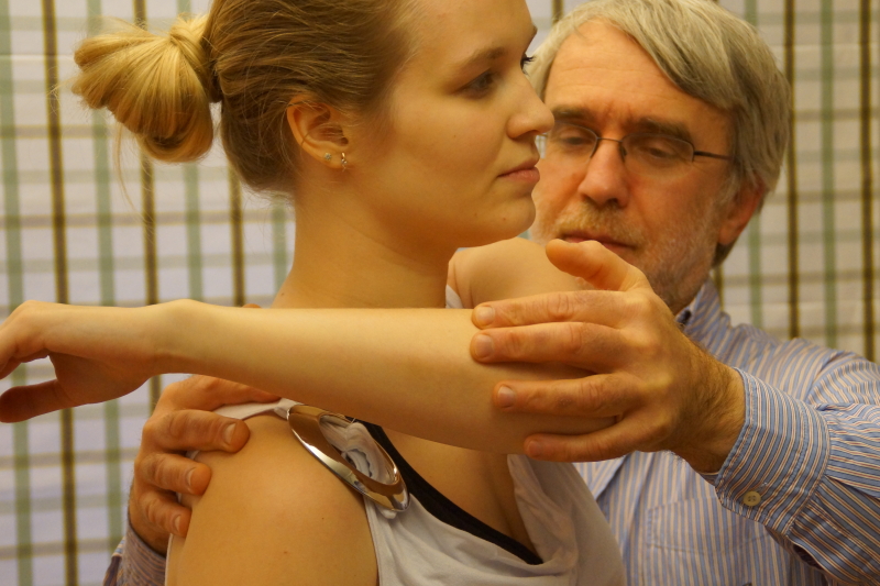
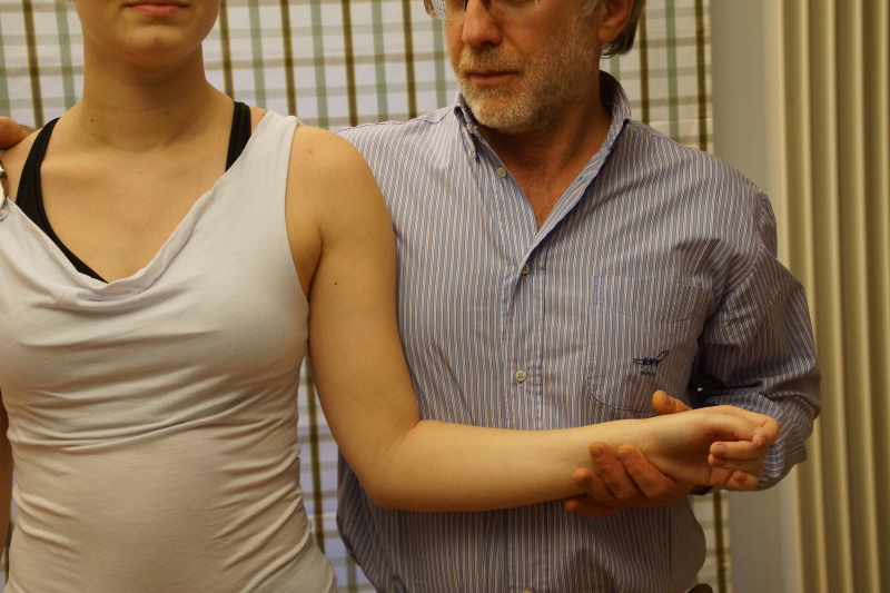 Passive lateral rotation
We assess pain, ROM and end-feel (elastic).
Make sure to block the heterolateral shoulder, so that the patient cannot do a compensatory movement, and sometimes (when the patient is very mobile) the therapist has to step slightly backwards to reach the real end of range.
This is important, since some lesions only cause end range pain.
In case of an arthritis, keep following detail in mind:
Stage III arthritis :
The lateral rotation movement will be quite painful resulting in a limitation with muscle spasm end feel due to increasing pain.
Stage I arthritis :
There will be limitation of ROM too, but only end range pain ; so, first we will find the limitation and when giving slight overpressure we will provoke some pain.
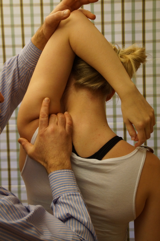
 Passive medial rotation
It is important to stabilize the patient’s arm, to prevent an improper execution of the test (we have to focus on medial rotation instead of retroflexion). We assess pain, ROM and end-feel (elastic).
A painful arc on medial rotation is possible, so, don’t stop the test movement too early! If it is painful, ask the patient where the pain is felt and try to continue the movement, allowing us to discover a possible painful arc.
A painful arc on medial rotation very often suggests the presence of a chronic subdeltoid bursitis.
The normal range on medial rotation is 90°. How do we assess a limitation ?
At this point in the examination we distinguish between a capsular and a non-capsular pattern.
Capsular pattern = limitation lateral rotation > abduction > medial rotation.
Note: the limitation of movement can be variable, but the ROM proportions between these three movements are important.
 Resisted adduction
We test : teres minor, maior, latissimus dorsi and pectoralis maior.
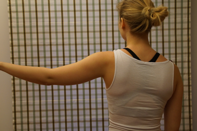
 Resisted abduction
We test the supraspinatus and deltoid.
 Resisted lateral rotation
We test the infraspinatus and teres minor.
Note the counter pressure at the patient’s heterolateral shoulder and the resistance given proximal from the wrist.
We ask the patient to do a lateral rotation, and perhaps it is useful to first show the patient what exactly we mean.
Our experience shows that many patients perform an abduction rather than a lateral rotation, which again endangers the final diagnosis.
 Resisted medial rotation
We test the subscapularis, teres maior, pectoralis maior and latissimus dorsi.
 Resisted elbow flexion
We focus on the biceps muscle.
 Resisted elbow extension
We focus on the triceps muscle.
Keep in mind that during a resisted test, inert structures may be compressed :
e.g. a resisted extension can compress an inflamed subdeltoid bursa, thus provoking pain into the C5-dermatome.
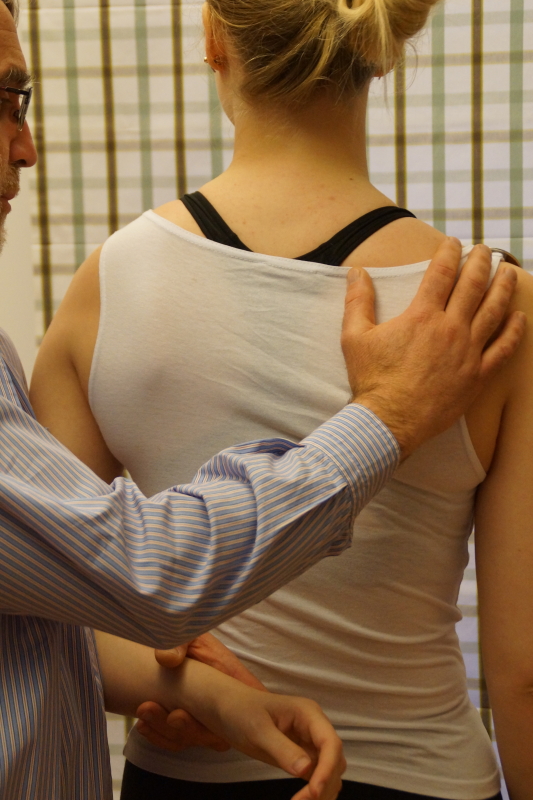
 Passive horizontal adduction
This test can be positive in three particular cases :
More accessory tests (e.g. in relation to mononeuritis) are illustrated in the film below.
An extensive overview of so-called impingement tests and it’s interpretation is illustrated in the powerpoint podcast below.