- How to diagnose and treat some ligament injuries in the knee?
- Ligament injury, specific history
- Clinical image of a ligamentous lesion
- Medial and lateral collateral ligament
- MCL clinical image acute lesion
- MCL treatment acute lesion
- MCL clinical image chronic lesion
- MCL treatment chronic lesion
- Lateral collateral ligament clinical image
- Coronary and cruciate ligaments
- Cross friction massage of the medial coronary ligament
- Cross friction massage of the medial collateral ligament
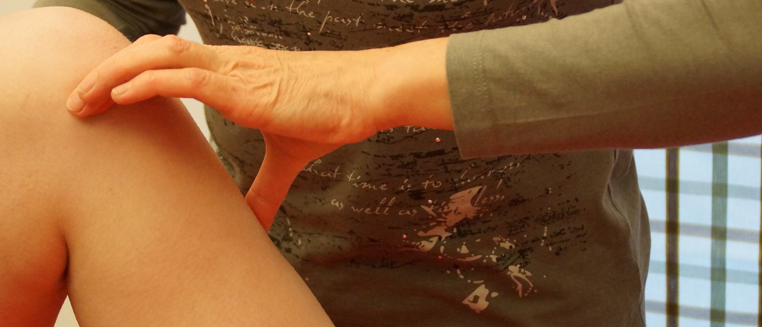
How to diagnose and treat ligament injuries in the knee
How to diagnose and treat some ligament injuries in the knee?
In fact it's quite easy to diagnose ligament injuries such as a collateral ligamentous lesion, or a sprain of a coronary ligament.
It's just a matter of correct interpretation of your functional examination.
Keep in mind that acute ligament injuries in the knee always show some kind of posttraumatic capsular reaction (swelling, capsular pattern), resulting in a clear limitation of movement of flexion and extension.
This will also have an impact on the treatment strategy.
Managing relevant clinical reasoning procedures are an essential tool to deal with musculoskeletal problems in an efficient and safe way. There's no need for "artificial hypercomplication".
Did you know that you can learn what really makes you a more successful therapist during the 5 days, world wide unique, Mastermind private training in orthopaedic medicine Cyriax?
Ligament injury, specific history
As already mentioned before, shortly after the injury the clinical pattern is dominated by the secondary traumatic arthritis, which makes it difficult to find the exact site of the initial lesion. Therefore, we try to discover in which direction the injury has taken place :
- was it a valgus strain with medial pain,
- or a varus strain with lateral pain,
- was it a flexion-rotation injury or
- did it happen in an unspecific direction and is the patient unable to tell us exactly what happened ?
An essential difference between the history of a ligament injury and that of a meniscal tear (bucket handle) is the immediate loss of function in a meniscal lesion, whereas the loss of function in a ligamentous lesion appears only after a few hours.
This means, for example, that a football player might continue the match when the lesion is purely ligamentous ; only after a few hours pain will increase together with swelling and limitation of range as a result of the post-traumatic capsular reaction.
Clinical image of a ligamentous lesion
The clinical pattern is different in the acute, subacute and chronic stage.
- The acute stage is characterized by the traumatic arthritis: warmth, fluid and a capsular pattern dominate the clinical pattern.
- In the subacute stage, the traumatic arthritis recedes gradually and the primary ligamentous test becomes more obvious: Valgus strain for the MCL, Varus strain for the LCL, Lateral rotation strain for the Medial coronary ligament and Medial rotation strain for the Lateral coronary ligament ; Lachman for the Anterior cruciate ligament and the Posterior drawer for the Posterior cruciate ligament.
- In the chronic stage the primary ligamentous test is positive ; the traumatic arthritis no longer exists. In the absence of treatment, the chronic stage is reached after about six weeks. With an early treatment by deep friction and mobilization, the lesion will not reach a chronic stage and clinical healing will occur after the subacute stage.
If the lesion is already in a chronic stage, then post traumatic adhesions may have formed.
Of course we hear the typical story of a chronic lesion (trauma – no active treatment – ADL OK – more intense activity not OK – some pain and swelling for some days – then OK again). Ligamentous adhesions can cause a slight limitation of passive movement.Medial and lateral collateral ligament
A lesion of the medial collateral ligament can occur on three different locations : - Most frequently at the jointline
- Or the tibial extent
- Or the femoral part
So, we always have to check three localisations by palpation.MCL clinical image acute lesion
- Clear capsular pattern with muscle spasm end feel
- Flexion more limited than extension
- P lateral rotation possibly slightly painful
- Valgus strain quite painful
MCL treatment acute lesion
- Progressive transverse friction massage from two positions: In as much flexion and in as much extension as possible
- First week daily treatment ; as from the second week about 3x/week
- Gentle active mobilization
- As from the subacute stage also proprioception.
MCL clinical image chronic lesion
- P flexion end range painful
- Possibly -10° limitation ROM
- P extension end range painful
- Possibly P lateral rotation end range painful
- Valgus strain painful
MCL treatment chronic lesion
- First treatment session o 10’ DTM in extension
- Manipulation in extension and lateral rotation
- 10’ DTM in flexion
- Manipulation in flexion
- Patient is advised to regularly move end range in flexion and extension
- As from the second session o 15’ of DTM 2-3x/week
- Proprioception.
Lateral collateral ligament clinical image
A lesion of the LCL looks clinically more moderate : there is less swelling and a minor capsular pattern in the acute stage, because of the fact that the LCL is not connected with the capsule. The main positive test will be the varus strain.Coronary and cruciate ligaments
The cruciate ligaments cannot be palpated of course. In some cases surgery is called for. In case of a medial coronary ligamentous lesion the main positive test will be the passive lateral rotation.
Also in an acute stage there will be a post-traumatic capsular reaction, which disappears in a chronic stage.
However, there is much less swelling in comparison to a lesion of the MCL. In a chronic stage we won’t be able to detect any limitation of passive movement. The coronary ligaments react well on cross friction massage
Cross friction massage of the medial coronary ligament
The starting position is 90° flexion and lateral rotation. For the left knee, we put the left index finger onto the tibial plateau. The finger nail faces upwards ; in this way, pressure can be applied in a downward direction. The thumb should be put far down, in order to maintain this pressure. The deep friction is the normal execution.
For a lateral coronary ligament, the knee is in medial rotation.
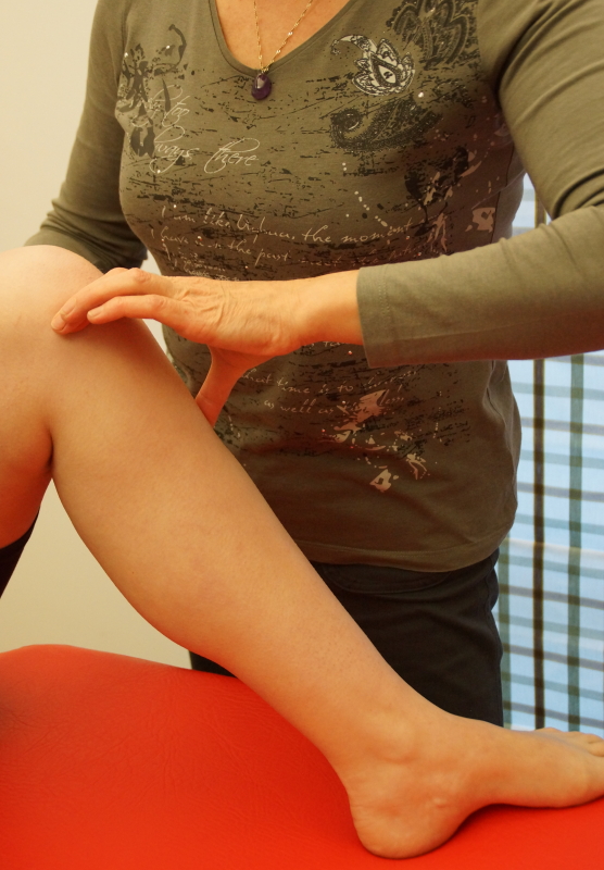 Cross friction massage for the medial coronary ligament
Cross friction massage of the medial collateral ligament
The friction is given in as much extension and flexion as possible. The knee is supported by a cushion. We locate the joint line and we palpate in a posterior direction until, beyond the midline, the ligament is found (a large flat structure). The deep friction is the normal execution.
For the deep friction in flexion, we find the lesion in extension first, keep our finger on it, and bend the knee. The deep friction, again, is the normal execution. Notice that the direction in which the ligament now lies has changed, and so has the direction of our friction.
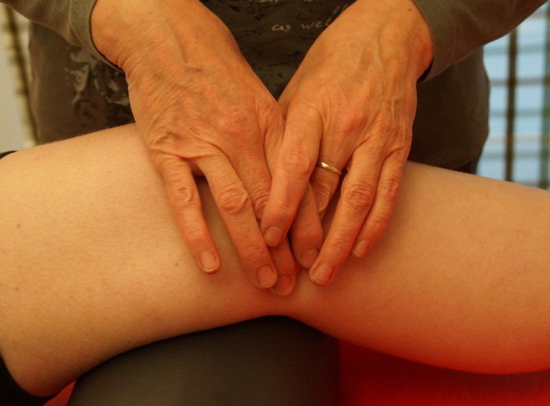 Cross friction massage of the medial collateral ligament (1)
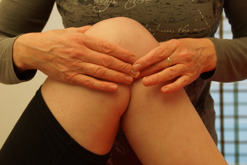 Cross friction massage medial collateral ligament (2)
