- How to interpret an orthopaedic physical examination of the extremities, based on Cyriax principles
- Introduction to the Cyriax orthopaedic physical examination
- A correct starting position
- Always ask neutral questions, never suggesting anything
- Distinction between a contractile and an inert lesion
- Inspection
- Cyriax orthopaedic functional examination
- What about the end feel?
- Lesion of an inert structure
- Lesion of a contractile structure
- Conclusion
- Multiple lesions – confusion ?

Orthopaedic physical examination, how to interpret?
How to interpret an orthopaedic physical examination of the extremities, based on Cyriax principles
It is very interesting to use some kind of standardized examination protocol when you examine soft tissue lesions of the upper or lower extremity.
Using a certain clinical reasoning algorythm fascilitates the diagnostic process. In the film below I would like to illustrate some of the "basics". Keep also in mind that the Cyriax clinical reasoning process in the extremities is quite different from the one used in the spine.
Introduction to the Cyriax orthopaedic physical examination
The Cyriax orthopaedic physical examination is a functional examination in which we use “selective tension” i.e. we test each structure separately or we test a group of structures.
First we would like to find the structure at fault, and then, if possible, the precise localization of the lesion in that structure. Therefore we sometimes need specific palpation in a specific structure. Sometimes palpation is obsolete since we may find a ‘localizing sign” in the functional examination, i.e. a specific positive tests which points us in the direction of the specific location where the lesion lies.
Note that the clinical reasoning strategy used in the extremities is different than the one used in the spine.
Please take into account certain rules when performing tests :
A correct starting position
Example :
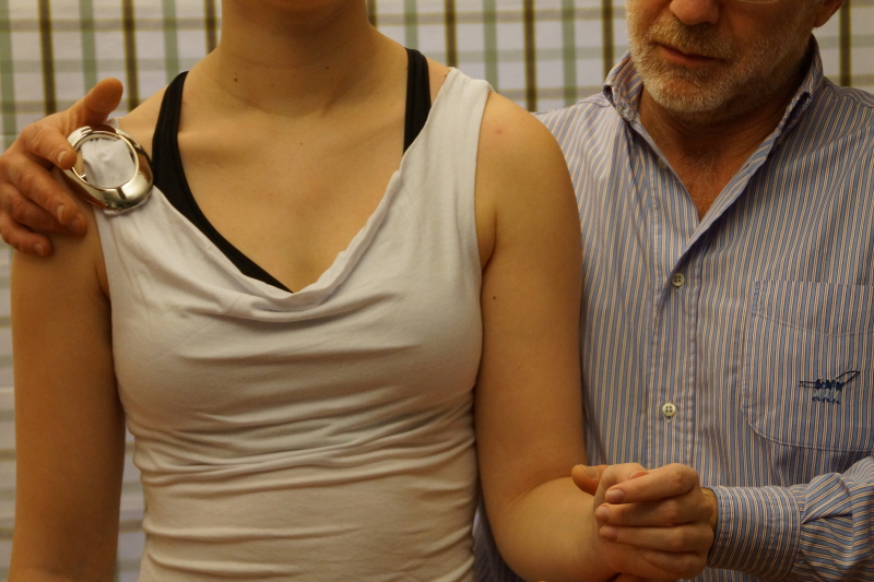
resisted lateral rotation in the shoulder. The heterolateral shoulder is stabilized and resistance is given at the distal part of the forearm, not at the wrist. In this way, we get a pure lateral rotation and not a shoulder abduction and wrist extension.
 Resisted lateral rotation in the shoulder
Always ask neutral questions, never suggesting anything
When performing the tests It is not a good idea to ask on each test “does that hurt?”.
This could be misleading and may easily lead towards an incorrect diagnosis.
Let’s imagine that the patient has some pain at rest, before doing any test : when we perform a test and ask him “does that hurt ?”, the answer obviously will be “yes”. But, in fact, the patient has the same pain during the test as the pain at rest. So, the test is negative.
We want to know whether a particular test has an influence upon his symptoms. A better question therefore would be : “does that change anything ?”.
The patient can give several answers :
- The pain increases
- The pain decreases
- No change
- Or, another pain is provoked somewhere else
- Example : a patient may suffer from lumbar pain and on performing an extension in standing he may provoke pain in the calf. This will have some diagnostic and therapeutic consequences.
- So, always ask where any pain is provoked !
I know...it's quite a challenge to manage to examine our patient and to reach a useful conclusion. The clinical reasoning procedure which has been proposed by Dr Cyriax is quite logical. Of course, time didn't stand still and we have to take into account interesting new scientific evolutions.
Wouldn't it be nice that you could save a lot of time by following a continuing education program that really meets your needs, that really makes you a better therapist? No irrelevant info, no "artificial hypercomplication", not too much (semi)scientific blablabla...
The perfect is solution is the Mastermind private training in modern orthopaedic medicine Cyriax.
OK, time for some more clinical reasoning...
Distinction between a contractile and an inert lesion
Typical for the clinical reasoning strategy in the extremities we would like to make a distinction between an inert or a contractile lesion.
Does the patient suffer from an inert and/or contractile lesion ? That’s the important clinical question.
Inert structures : capsule, disc, meniscus, ligament, fascia, dura mater, nerves, bursa.
Contractile structures : muscle belly, body of tendon, musculotendinous junction, and tenoperiosteal junction.
A clinical examination is composed of four main parts : history, inspection, functional examination and, if necessary, palpation.
Palpation can be very useful in the extremities ; in the spine, however, palpation for pain and mobility is often very misleading and mostly irrelevant !
Inspection
Inspection not only includes the interpretation of e.g. redness, swelling, antalgic posture, wasting, etc. It also means that we have to observe the patient, his daily behaviour, the way he sits, stands, the expression on his face. This could be very useful to detect certain inherent unlikelihoods.
Example :
a patient states that she cannot bend forwards because of an excruiating pain ; however, when seated for some time during the history-taking, she manages to bend quite normally to pick up something from her handbag.
Cyriax orthopaedic functional examination
There is a difference in the strategy for what is concerned the extremities and the spine. In contrast to the spine, in the extremities we practically never use active test movements.
Why ? During an active movement both contractile and inert structures are “in action”. So, if a patient states he has pain or limitation on performing an active movement we cannot make the clinical distinction between a lesion of a contractile or an inert structure. Therefore active testmovements are not very useful in the extremities.
An active test movement, however, provides us some information on the patient’s willingness to perform the movement. Sometimes this could be useful to discover a simulating patient.
We prefer to concentrate more on the use of passive and resisted movements.
>>> Passive test movements
Passive tests are performed until we reach the real end of the movement.
Those tests are the main tests for the integrity of the intert structures.
We have to test three variables :
- Pain
- Range of motion
- End feel
>>> Resisted test movements
Resisted tests are performed from a neutral postion (as less tension as possible on inert structures) ; we ask for a maximal isometrical contraction.
Those tests are the main tests for the integrity of the contractile structures.
We have to test two variables :
- Pain
- Force
Of course, a passive test can also put strain on contractile structures and a resisted test could compress some inert structures. This is a challenge we face during the interpretation of the test results.
On performing a clinical examination, always ask yourself what exactly you are testing and which variables you are testing.
Also always question the validity of any test : does the test have a good intertester or intratester reliability ; is there a relevant specificity or sensitivity of the test ? Try to get as much information as possible by using a less tests as possible.
There really is no need for any “artificial hypercomplication”. Unfortunately many tests, frequently used in general manual therapy, have a very poor validity, therefore making them rather obsolete.
>>> Localizing signs
Sometimes we find certain positive tests in the examination which help us to determine the specific location of the lesion.
Example :
A shoulder patient has pain on resisted abduction, pointing in the direction of a supraspinatus or a deltoid lesion. We also find a painful arc (i.e. a painful passage between two painfree passages during elevation).
The painful arc suggests something is temporarily squeezed in between humerus and acromion, thus excluding the deltoid, but pointing in the direction of the tenoperiosteal part of the supraspinatus.
A painful arc in the shoulder is a common clinical finding and could be related to a number of lesions.
>>> Palpation
At rest we could feel warmth, swelling and synovial thickening. During a movement we also could feel the end feel or some crepitation.
Once we determined the structure at fault, but we don’t know the exact localisation of the lesion yet, then of course we need to palpate for pain in this specific structure.
What about the end feel?
On passive testing, we go for the end of the movement and then we slightly provoke at end range in order to test the specific end feel. There are normal and pathological end feels. Please don’t use any force, but merely perform a gentle provocation.
>>> Normal / physiological end feel
- Hard : e.g. elbow extension, knee extension
- Capsular (elastic) : e.g. rotations at shoulder, elbow, hip
- Extra-articular (tissue approximation) : flexion at elbow, hip.
>>> Pathological end feel
- Too hard : e.g. osteoarthrosis
- Too soft : e.g. loose body in the elbow joint, extension has a softer end feel instead of hard
- Muscle spasm (involuntary or voluntary muscle contraction) : e.g. arthritis
- Springy block : e.g. meniscus subluxation causing a limitation of knee extension.
Lesion of an inert structure
A lesion of an inert structure is brought in mind when one or more passive tests are positive, while all the resisted tests are negative .
The question however is, which inert structure is at fault ?
We will make a distinction between the joint capsule and the other inert structures ; therefore we need to interpret the findings.
>>> Capsular pattern ?
The capsular pattern is a combination of pain and/or limitation, which points in the direction of a joint problem.
Each joint, under muscular control, has a specific capsular pattern.
Example :
- Capsular pattern in the shoulder is a limitation of certain passive movements : lateral rotation > elevation > medial rotation.
- Capsular pattern of the elbow : flexion is more limited than extension.
When a capsular pattern has been found on examination, then this had very clear clinical implications : or the patient suffers from osteoarthrosis or from some sort of arthritis.
The arthritis might be minor or severe, the limitation therefore could be slight or
considerable ; the proportions, however, remain always the same.
If the end feel on passive movement is harder, we first think of osteoarthrosis. When we find a muscle spasm end feel, we think of an arthritis i.e. an active inflammatory lesion.
>>> Non-capsular pattern
The non-capsular pattern is any other combination of pain and/or limitation of movement than the one found in case of a capsular pattern.
The lesion is not an arthritis or an arthrosis but something else.
Example :
>>> Ligamentous adhesions
After an injury, adhesions may interfere with the normal range of movement. We then expect a very typical history, together with a typical clinical image :
ONE movement is SLIGHTLY limited by LOCALIZED pain.
History : there has been an injury followed by only immobilization or rest. After some time the structure functions relatively normal for activities of daily life, but doesn’t function optimal during and after more intense activities. There will be some after pain and perhaps some swelling for a few days, after which symptoms spontaneously disappear again.
>>> Internal derangement
A joint is blocked in one direction and free in another direction . E.g. meniscus subluxation at the knee, acute torticollis or an acute lumbago.
>>> Extra-articular limitation
There will be GROSS limitation in ONE direction only, with normal movement in all other directions. E.g. passive knee flexion is markedly limited in case of an acute lesion in the quadriceps muscle belly (because of muscle spasm).
Lesion of a contractile structure
One or more positive resisted tests (and passive tests being negative) suggest a lesion of a contractile structure.
Although a positive resisted test can also be related to other problems such as :
- A fracture
- Compression of an inert structure (e.g. bursitis)
- Psychogenic disorders
- Metastases.
The most obvious clinical image is one positive resisted test, while all the other passive
and resisted tests are negative. (Although a passive test can stretche or pinche the affected structure).
Several muscles do have a double function, therefore the differentiation is made by means of complementary tests.
Always remember we test two variables : a resisted test may be painful and/or weak. This leads to a number of interpretations :
| Pain | strength |
|
1 | + | - | This is a minor muscular or tendinous lesion
|
2 | - | weakness | Could be the result of a nervous lesion (mononeuritis, compression nerve root) or a complete tendinous rupture
|
3 | + | weakness | Or this is a severe lesion (fracture, cancer) or, more frequently is the result of a partial tendinous rupture
|
4 | All tests + | - | Or the patient is hypersensitive or suffers from a psychogenic problem
|
Conclusion
At the end of the Cyriax orthopaedic physical examination, an accurate assessment of the findings is needed. During the clinical reasoning process we have to establish whether the patient has a lesion of an inert and/or contractile structure.
If possible we dertemine the structure at fault, but perhaps we don’t know yet from where exactly in this structure the problems arises, therefore we will need a selective palpation for tenderness.
Keep also in mind to concentrate at all times on relevant functional examination procedures and to abort irrelevant testing.
Unfortunately therapists often claim to “feel” e.g. hyper- and hypomobilities which can’t be discovered in a reliable way. So, avoid to “think” that you “feel” something.
Keep in mind that palpation for tenderness is more useful in joints such as knee, elbow, hand and foot and that palpation for tenderness could be quite misleading in the spine. Actually, palpation for tenderness in relation to spinal problems is very often irrelevant.
When, after the history and the clinical examination, we have reached a presumable diagnosis, there is still room for error. If necessary a presumable diagnosis can be doublechecked by means of a specific local anaesthetic infiltration or a diagnostic friction massage.
Compare the outcome of the positive test(s) before and after the application of infiltration or friction massage. This system can be very useful in the extremities.
It is an illusion to think that we are always going to be able to reach a solid diagnosis.
>>> Minor problem ?
Perhaps the problem is just a minor problem and there is just some slight pain in certain circumstances. Solution : repeat tests, increase the frequency, increase the intensity ; try to provoke the patient’s symptoms. Remember, there always should be a link between the symptoms and certain positions and/or activities.
>>> Too acute ?
We may find too many positive tests or we can’t perform some tests because it is too painful, which makes the assessment more difficult. In such a case, the history becomes much more important. It is adviseable to repeat the examination on another occasion.
>>> Lesion outside the sphere of orthopaedic medicine ?
Take into account e.g. a visceral lesion, which is not obvious in a basic orthopaedic evaluation. It is important to interpret inherent (un)likelihoods from the history and the examination !
Multiple lesions – confusion ?
A patient suffers from a multiple or double lesion. Most likely this won’t be obvious during the first clinical examination. Therefore stick to following hints :
- treat the easiest lesion first
- treat the most frequent lesion first
- treat the most obvious lesion first
- treat the most painful lesion first.
During the treatment of the first lesion, the clinical image of that lesion will improve ; on re-testing then the image of the second, previously more “hidden” lesion, will become more prominent.
So, re-test on several occasions.
“Treat what you understand and leave alone what you don’t understand.”
